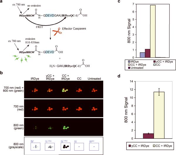
Figure 1. Fluorescence can be detected in oocytes microinjected with the IRDye and cytochrome c.
(A) Schematic of the LI-COR IRDye® 800CW/QC-1 CSP-3, which contains a non-fluorescent quencher dye (IRDyeQC-1) conjugated to a donor dye (IRDye800CW) by a DEVD-containing peptide. Proteolytic cleavage by active effector caspases separates the dyes, abolishing fluorescence resonance energy transfer (FRET) thereby restoring the donor dye fluorescence. Adapted with permission from LI-COR® Biosciences, Inc. (B) Oocytes were injected as indicated and imaged 30 minutes later. yCC, yeast cytochrome c, and CC, cytochrome c, both injected at 67 nM. (C) Graphical representation of the total quantitated signal in the 800 nm channel for each collective group of oocytes from B. Signal from individual oocytes in B can also be measured to calculate the average oocyte fluorescence (Supplemental Figure 1a and 1b). (D) Total quantitated signal in the 800 nm channel for each collective group of oocytes was averaged from independent microinjection experiments (n=21) of IRDye with either yCC or CC and is displayed +/− S.E.M.

