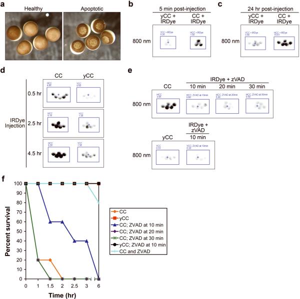
Figure 3. Caspases are rapidly activated in response to cytochrome c; caspases remain active for hours although their activity is only required for 10 minutes to ensure apoptosis.
(A) Oocytes were injected with yCC (healthy) or CC (apoptotic) at 106 nM, and photographed 3 hours later. (B) Oocytes microinjected with IRDye and either yCC or CC at 67 nM were imaged for fluorescence after 5 minutes, demonstrating rapidity of the signal in response to even small amounts of CC. Average oocyte fluorescence is displayed in Supplemental Figure 1e. (C) Oocytes microinjected with IRDye and either yCC or CC at 533 nM were imaged after 24 hours, demonstrating a stable, selective induction of signal in CC-injected oocytes even at high yCC concentrations. Average oocyte fluorescence is displayed in Supplemental Figure 1f. (D) Oocytes microinjected with either CC or yCC at 80 nM at t=0 were then injected with IRDye at various times indicated, and imaged 30 minutes later. Average oocyte fluorescence is displayed in Supplemental Figure 1g. (E) CC or yCC was microinjected at 533 nM at t=0, then IRDye and z-VAD-fmk (70 nM) were injected at times indicated and then imaged 30 minutes later. Note z-VAD-fmk-mediated signal inhibition even at high concentrations of CC. Average oocyte fluorescence is displayed in Supplemental Figure 1h. (F) Survival curve based on observation of apoptotic morphology of oocytes that had been treated as described in E.

