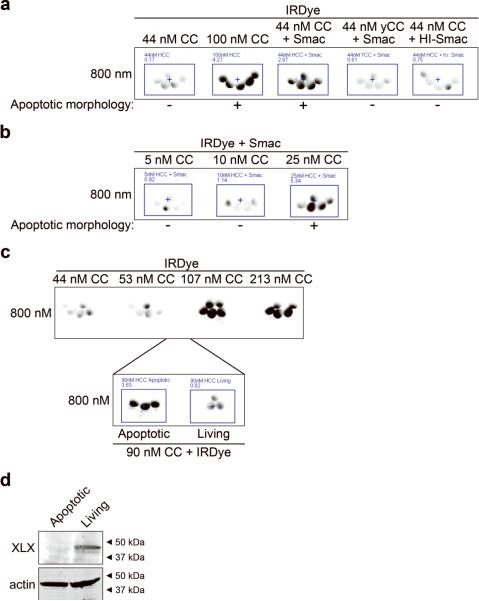
Figure 4. A cytochrome c threshold exists in oocytes and is lowered by Smac addition.
(A) Oocytes were microinjected with IRDye and either sub-threshold CC (44 nM), supra-threshold level of CC (107 nM), 44nM CC plus Smac protein, 44nM yCC plus Smac, or 44nM CC plus Smac that had been heat-inactivated by heating at 95°C for 5 minutes (HI-Smac) and then imaged after 40 minutes. Smac was injected to a concentration of 280 nM. Average oocyte fluorescence is displayed in Supplemental Figure 1i. (B) Low doses of CC were injected with the IRDye and Smac and imaged after 30 minutes. Average oocyte fluorescence is displayed in Supplemental Figure 1j. (C) Oocytes were injected with IRDye and increasing doses of CC and imaged after 45 minutes. Naive oocytes from the same batch were then injected with IRDye and 90nM CC, observed for apoptotic morphology and then 3 apoptotic and 3 living oocytes were imaged for fluorescence. Average oocyte fluorescence is displayed in Supplemental Figure 1k. (D) Lysates were prepared from the apoptotic and living oocyte groups described in C and then immunoblotted for XLX.

