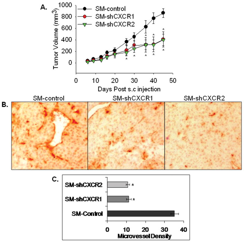
Figure 2. knock-down of CXCR1 or CXCR2 reduces melanoma tumor growth and decreases microvessel density in vivo.
Melanoma cells (SM-shCXCR1, SM-shCXCR2 or SM-control) were subcutaneously (s.c.) injected into the right flank region of nude mice. Tumor volume was measured twice weekly with a caliper and was calculated by using the formula π/6 × (smaller diameter) 2 × (larger diameter). A, Tumor volume from day 0 to day 45 (n=6). Growth of SM-shCXCR1 and SM-shCXCR2 were significantly reduced (p<0.05) compared with SM-control group. B, Immunohistochemical staining for microvessels with anti-GS-IB4. The representative pictures are shown at 200× C, The values are average number of microvessels ± SEM. Microvessel density was quantitated microscopically with a 5×5 reticle grid at 400× magnification. The values are mean ± standard error of mean (SEM). *Significantly different from SM-control (p<0.05).

