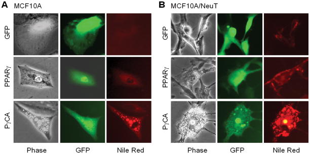
Figure 1. PγCA induces lipid formation of MECs cells transformed by ErbB2 oncogene.
MCF10A or MCF10A/NeuT cells were infected with retroviral vector expressing GFP, PPARγ, and PγCA. 48 hr post-infection, cells were stained with Nile Red (100 ng/ml). Images were taken under a florescent microscope. GFP-positive cells were analyzed for accumulation of oil droplets within the cells as shown by Nile Red staining.

