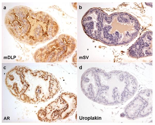
Figure 1. Expression of markers in mouse dorsolateral prostate (DLP).
Control mouse seminal vesicle immunohistochemistry for dorsal lateral prostate secretions (mDLP), seminal vesicle secretions (mSV), androgen receptor (AR) and uroplakin. As expected mouse DLP shows cytoplasmic brown staining in the luminal epithelial cells with antibodies raised against mDLP secretions (a) but not with those raised against mouse seminal vesicle secretions (b) or uroplakin (d). Nuclear brown staining is seen in both epithelial and some stromal cells when antibodies against AR were used (c)

