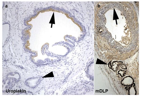
Figure 3. Development of prostatic tissue.
Recombinant graft of trigone epithelium and urogenital sinus mesenchyme demonstrating the development of prostatic structure from urothelium under the influence of urogenital sinus mesenchyme. Fragments of the grafted urothelium can be seen (arrows)continuing to express uroplakin (a). More characteristic prostatic glandular epithelium grows from these structures (arrowheads) under the influence of urogenital sinus mesenchyme (as previously described) these structures show positive staining using antibodies targeting dorsal lateral prostate secretions (mDLP) (b).

