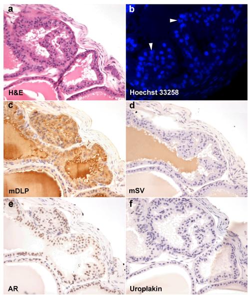
Figure 4. Tissue recombinant composed of mouse trigone epithelium and rat urogenital sinus mesenchyme.
Recombinant graft of trigone epithelium and urogenital sinus mesenchyme stained with H&E (a), Hoechst 33258 (b), mDLP (c), mSV (d), AR (e), and uroplakin (f). Clear glandular epithelial differentiation with columnar secretory luminal cells is seen. Epithelial nuclei stained with Hoechst 33258 (b) demonstrates the speckled nuclear patterning characteristic of mouse chromatin packaging (white arrowheads) in epithelial cells while the stromal cells show the more diffuse staining characteristic of rat nuclei. Recombinant glandular structures show brown cytoplasmic staining in luminal epithelial cells for mDLP (c) not mSV (d) or uroplakin (f). Brown nuclear staining is seen using antibodies against AR (e).

