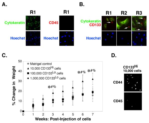
FIGURE 1. Identification of a CD133-hi tumorigenic sub-population of carcinoma cells in patient ascites.
A,B. Unsorted ovarian carcinoma cells from patients R1, R2 or R3 were plated on chamber slides for 24h and processed for immuno-staining as indicated. Cells were visualized by epifluorescence. Arrows in B indicate CD133-hi cells. C, D. Carcinoma cells from patient R1, magnetically sorted for CD133, were injected into the peritoneum of SCID mice as indicated. C. Tumor growth after injection of 104 CD133-hi cells in Matrigel was compared to tumor growth after injection of 105 or 106 CD133-lo cells in Matrigel (or Matrigel alone, as control), as assessed by percent change in weight due to ascites accumulation [(weight at week n/weight at day 0)-1]*100. Significant differences were found between CD133-hi @ 104 cells and: CD133-lo @ 105 cells (*p<.05), CD133-lo @ 106 cells (#p<.05), or Matrigel control (%p<.05), as determined by one-way ANOVA with Bonferroni comparison of significant differences between points. D. After 7 weeks, ascites was drawn from the peritoneum of animals receiving CD133-hi cells and cells plated for 24h before staining for CD44 and CD45. Ascites was absent from animals that received CD133-lo cells or Matrigel alone and significant numbers of cells could not be retrieved from these animals.

