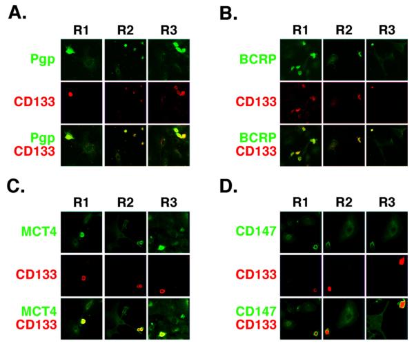
FIGURE 2. Co-distribution of signaling and transporter proteins in CD133-hi primary ovarian carcinoma cells.
A-D. Low power confocal images of unsorted ovarian carcinoma cells from patients R1, R2 and R3 that were plated on chamber slides for 24h and stained for CD133 (red) and either Pgp (A), BCRP (B), MCT4 (C), or emmprin/CD147 (D) (green). Most cells lacked significant staining for CD133 (CD133-lo cells); these cells showed relatively low levels of Pgp, BCRP, MCT4 and emmprin. A few cells stained brightly for CD133 (CD133-hi cells); these cells also stained strongly for Pgp, BCRP, MCT4 and emmprin. Similar results were obtained with EGFR and CD44 (see Fig. 5A), as well as with ErbB2 and MCT1 (not shown).

