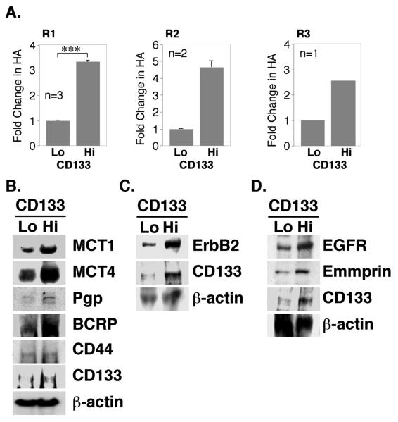
FIGURE 3. Elevated expression of hyaluronan, signaling proteins and transporter proteins in CD133-hi primary ovarian carcinoma cells.
A. Ovarian carcinoma cells from patients R1, R2 and R3, magnetically sorted for CD133, were plated in 6-well dishes for 24 hours. Conditioned media were collected and hyaluronan quantified by an ELISA-like assay. Results were normalized to cell number. For R1 cells, error bars express standard deviation in the mean of three separately sorted batches; significant differences (***p<.001) between CD133-lo and CD133-hi cells were observed. For R2 cells, error bars represent range of two measurements. Due to limitations in amounts of primary material, the statistical significance of differences could not be determined in R2 and R3 cells since n<3; however, the differences obtained were large. B-D. Ovarian carcinoma cells from patient R1, magnetically sorted for CD133, were plated in 6-well dishes for 24h. Western blot analysis of whole cell lysates (50μg/lane) demonstrated elevated expression of CD133, MCT1, MCT4, Pgp, BCRP, ErbB2, EGFR, and emmprin in CD133-hi cells. Expression of CD44 (B) was approximately equivalent in CD133-hi and CD133-lo cell lysates. Panel B shows a blot obtained with lysates from one batch of sorted cells, whereas panels C and D show two separate blots obtained with lysates from a second sort.

