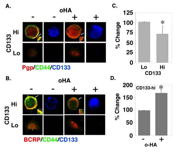
FIGURE 4. Co-localization of drug transporters with CD44 in CD133-hi cells and disruption by hyaluronan oligomer treatment.
A, B. Unsorted ovarian carcinoma cells from patient R1 were plated on chamber slides for 24h, treated with and without 100 μg/ml hyaluronan oligomers for 1 hour, and then processed for immuno-staining. Cells were stained for CD44 (green), for Pgp (A) or BCRP (B) (red), and for CD133 in order to distinguish CD133-hi and CD133-lo cells (blue; shown separately). CD133-hi and CD133-lo cells were visualized by confocal microscopy at a z-plane corresponding to the approximate center of the cell. Note co-localization (yellow) of CD44 and transporters at the plasma membrane of untreated CD133-hi cells, and internalization of Pgp (A) and BCRP (B) in hyaluronan oligomer-treated CD133-hi cells. Arrows indicate areas of the plasma membrane and adjacent cytoplasm shown at higher magnification in the insets. C, D. Ovarian carcinoma cells from patient R1, magnetically sorted for CD133, were treated with and without 100 μg/ml hyaluronan oligomers for 1 hour in feed medium containing 2.5 μM FURA 2-AM, and analyzed for fluorescence as described in Methods. CD133-hi cells showed higher efflux activity than CD133-lo cells (C); the hyaluronan oligomers inhibited efflux in the CD133-hi cells (D). Error bars express standard deviation in the mean of triplicate wells. Significant differences were observed (*p<.05) as determined by Student's t-test. The results are representative of three or more independent experiments.

