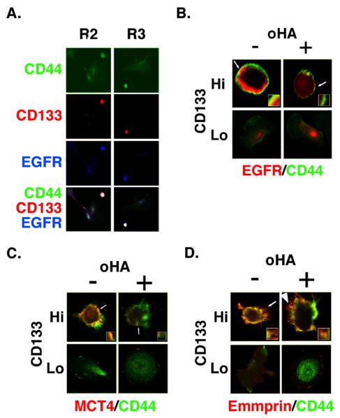
FIGURE 5. Co-localization of EGFR and MCT4 with CD44 in CD133-hi cells and disruption by hyaluronan oligomer treatment.
A. Low power confocal images of unsorted ovarian carcinoma cells from patients R2 and R3 that were plated on chamber slides for 24h and stained for CD44, CD133, and EGFR. CD133-lo cells, which comprised the majority of cells, showed dispersed staining for CD44 and low levels of EGFR. CD133-hi cells stained strongly for CD44 and EGFR. B-D. Higher power images of unsorted ovarian carcinoma cells from patient R1, that were plated on chamber slides for 24h, treated with and without 100 μg/ml hyaluronan oligomers for 1 hour, and then processed for immuno-staining. Cells were stained for CD44 (green), for EGFR (B), MCT4 (C) or emmprin (D) (red), and for CD133 in order to distinguish CD133-hi and CD133-lo cells (not shown). CD133-hi and CD133-lo cells were visualized by confocal microscopy at a z-plane corresponding to the approximate center of the cell. Note co-localization (yellow) of CD44 with EGFR, MCT4 and emmprin at the plasma membrane of untreated CD133-hi cells and internalization of EGFR (B) and MCT4 (C), but not emmprin (D), in hyaluronan oligomer-treated CD133-hi cells. Similar results were obtained for MCT1 and ErbB2 (not shown). Note that internalization of MCT4 is difficult to visualize here due to dispersion throughout the cytoplasm, but is clearly visible on increasing gain (e.g. see (21)). Increased gain did not reveal intracellular emmprin. Arrows indicate areas of the plasma membrane and adjacent cytoplasm shown at higher magnification in the insets.

