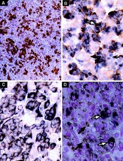
Figure 2.
The HSI,IIhGH transgene was expressed in somatotropes but silenced in lactotropes. (A) Representative section of a pituitary from an HSI,IIhGH (Fig. 1E) adult transgenic mouse stained with mAb9. The number and distribution of mAb9-positive cells (brown cytoplasm) corresponded to that seen with the −40hGH transgene (Fig. 1A) but the staining is more intense because of the higher level of transgene expression. (B) Following double immunostaining, mAb9 positivity (brown) was detected only in the cytoplasm of cells also positive with anti-Pit-1 (blue nuclei). A representative pituitary cell positive for both Pit-1 and hGH (white arrow), another pituitary cell-type positive for Pit-1 but not hGH (barbed black arrow), and a cell negative for both (black arrow) are indicated. (C) Double staining colocalized mGH (brown) and hGH (mAb9; charcoal black) in the cytoplasm of the same cells, yielding a dirty brown reaction product (compare with the pure brown reaction product in A). (D) Double staining was also carried out with mAb9 (charcoal black diffuse cytoplasmic stain; white arrows) and anti-Prl (granular rust-colored stain with juxtanuclear Golgi pattern of reactivity; black arrows). Mature lactotropes that expressed mPrl but not hGH, indicating appropriate silencing of the HSI,IIhGH transgene, were clearly identifiable (black arrow) and were distinct from cells expressing hGH (white arrow). (A and C were counterstained with hematoxylin, B was not counterstained, and in D the nuclei were counterstained with nuclear fast red. A, ×70; B–D, ×170.)

