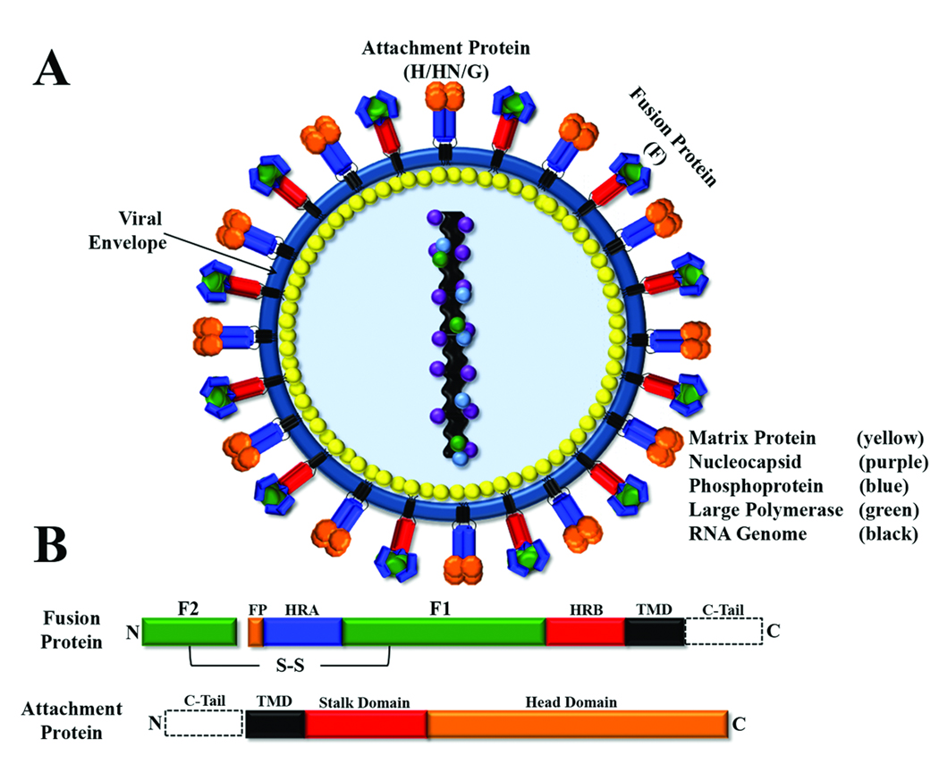
Figure 1. Schematic of paramyxovirus virion and surface glycoproteins.
A) Schematic of a paramyxovirus; viral membrane shown in blue. B) Conserved domains of paramyxovirus fusion and attachment proteins. Domain abbreviations: fusion peptide (FP, orange); heptad repeat A (HRA, blue); heptad repeat B (HRB, red); transmembrane domain (TMD, black); cytoplasmic tail (C-Tail, dotted box); disulfide bond (S-S).

