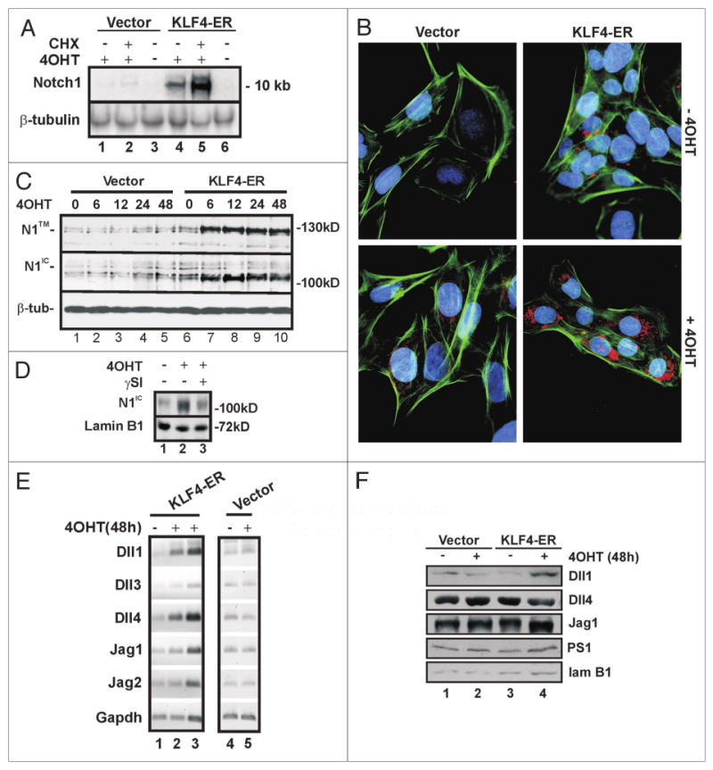
Figure 1.

KLF4 induces Notch1 expression in RK3E cells. (A) Notch1 is induced by KLF4 in RK3E cells without de novo protein synthesis. KLF4-ER cells or vector cells were treated with 4OHT +/- CHX for 2 h. Notch1 and -tubulin (loading control) mRNAs were analyzed by northern blot. (B) KLF4-ER cells or vector cells were induced with 4OHT for 10 h. Notch1 expression (red) was analyzed by immunoflouresence using N1-C20 antibody, which primarily detects cytoplasmic Notch1. DAPI (blue) and phalloidin (green) stain the nucleus and cytoskeleton, respectively. (C) Vector or KLF4-ER cells were treated with 4OHT for the number of hours indicated above the lanes. Notch1 expression was analyzed by immunoblot using antibodies N1-C20 (top) and N1-Nter (center) which predominantly recognize N1™ and N1IC respectively. -tubulin (bottom) served as a loading control. Molecular weight markers are indicated on the right. (D) KLF4-ER cells were treated with 4OHT or vehicle for 48 h. To test for a role of -secretase, cells were treated with SI or vehicle for 3 h prior to preparation of extract. Immunoblots were probed with N1IC-Nter and lamin B1 (loading control) antibodies. (E) Semi-quantitative RT-PCR analysis was performed using total RNA isolated from Vector or KLF4-ER-cells treated with 4OHT for 48 h. To demonstrate that PCR reactions were not saturated, lane 3 contained twice as much input RNA as lane 2. Gapdh served as control. (F) Immunoblot analysis of Notch pathway genes. Lamin B1 served as a loading control. Cells were treated as indicated for 48 h.
