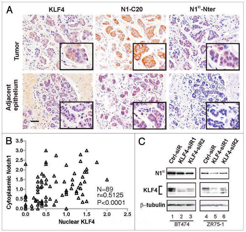
Figure 2.

Co-expression of KLF4 and Notch1 in human breast tumors. (A) Subjacent sections of a primary human breast tumor were stained with the indicated antibodies. Within the section were areas representing either infiltrating ductal carcinoma (Tumor, upper row) or adjacent, uninvolved epithelium (lower row). Scale bar, 50 μm. (B) Pearson correlation analysis of KLF4 and N1-C20 immunostaining in 89 cases of breast cancer. Tumors with the same scores appear as one data point. (C) Immunoblot analysis of N1IC following siRNA-mediated suppression of KLF4 in BT474 and ZR75-1 human breast cancer cell lines. The upper and lower portions of a filter representing a single SDS-PAGE gel were queried in parallel with N1IC-Nter or KLF4 (H180) antibodies, respectively. The lower portion was reprobed with β-tubulin antibody (loading control).
