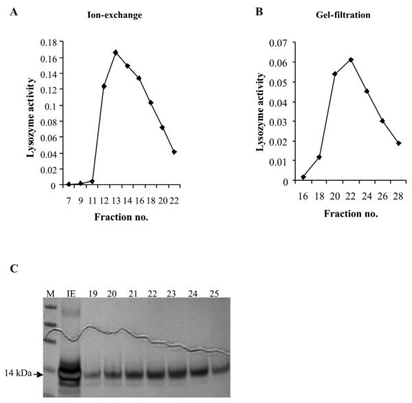
Figure 2.
Purification of lyoszyme c-1 from conditioned medium of 4a3B cells. A. Ion-exchange chromatography of the crude cell culture medium. B. Gel filtration chromatography of peak fractions from A. on Sephadex G-75. C. Silver stained gel from ion-exchange peak and G-75 fractions; Lane IE- ion exchange peak, lanes marked 19-25 are the respective fractions from B. Fraction 22-24 were used for characterization of activity under varied conditions. Lysozyme band is seen at an apparent molecular mass of 14 kDa marked by an arrow.

