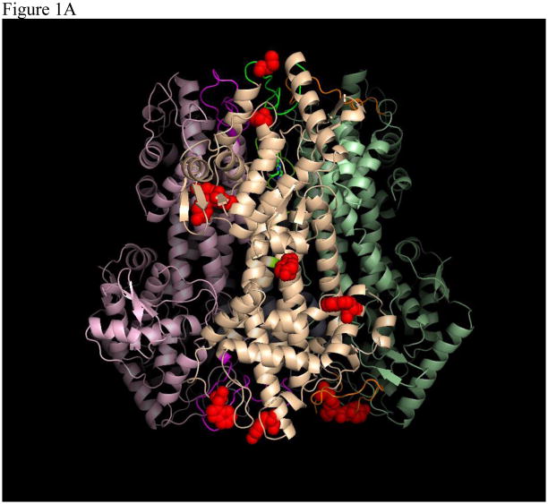
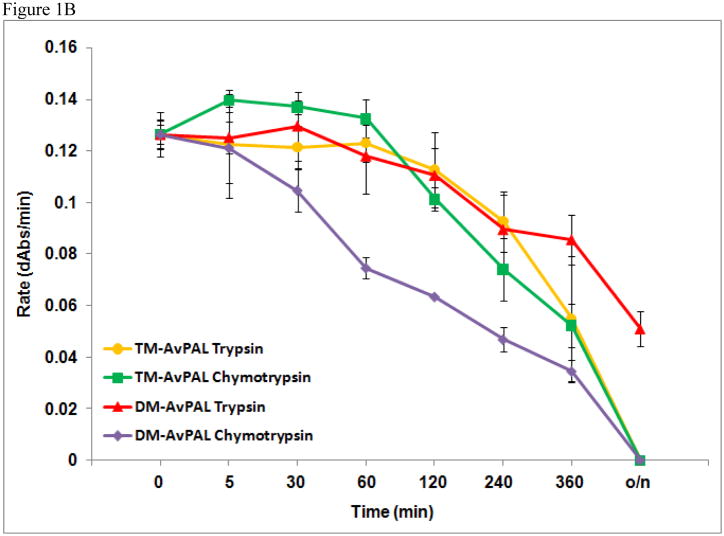
Figure 1.
(A) Tetrameric structure of AvPAL (PDB 2NYN). Surface-exposed potential trypsin and chymotrypsin cleavage sites on one out of the four monomers are represented by red spheres. (B) Comparison of protease resistance between TM-AvPAL and DMAvPAL incubated with chymotrypsin or trypsin (40 μg/ml) solutions at pH 8.0. The various plots were normalized at time zero to facilitate comparison.


