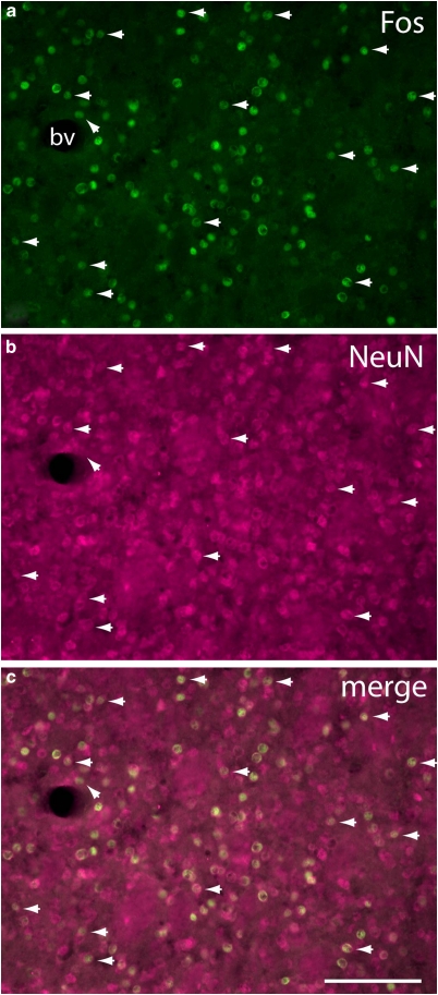
Figure 4.
Fluorescence micrographs of the central caudate-putamen (see Figure 2d) of an SAC 1 day rat illustrating localization of Fos-immunoreactive nuclei (a) within cells identified in panel (b) as neurons on the basis of immunoreactivity against the neuronal nuclear antigen (NeuN). An obliquely oriented arrowhead near a blood vessel (bv) marks the only Fos immunoreactive structure in the field not colocalized with a NeuN immunoreactive structure. Horizontally oriented arrowheads mark examples illustrating that NeuN immunoreactive structures, because NeuN often stains both the cell body and nucleus, are usually larger than the colocalized Fos-labeling. The disparity in the size and shape of co-localized Fos- and NeuN-immunoreactive structures, also apparent in the merged image (c) rules out the possibility that the near complete assocation of Fos-ir nuclei with NeuN structures is due to bleed-through of the FITC signal. Scale bar: 100 μm.

