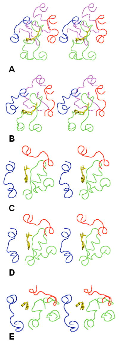
Figure 1.
Stereoviews of the X-ray structures of the EC loops in five GPCRs shown as lines in red (EC1), green (EC2), blue (EC3) and magenta (the N-terminal tail). Ligands crystallized with the GPCRs are shown as sticks in yellow. A) bRh; B) sRh; C) β2AR; D) β1AR; E) A2AR. The view is from the extracellular space normal to the membrane plane.

