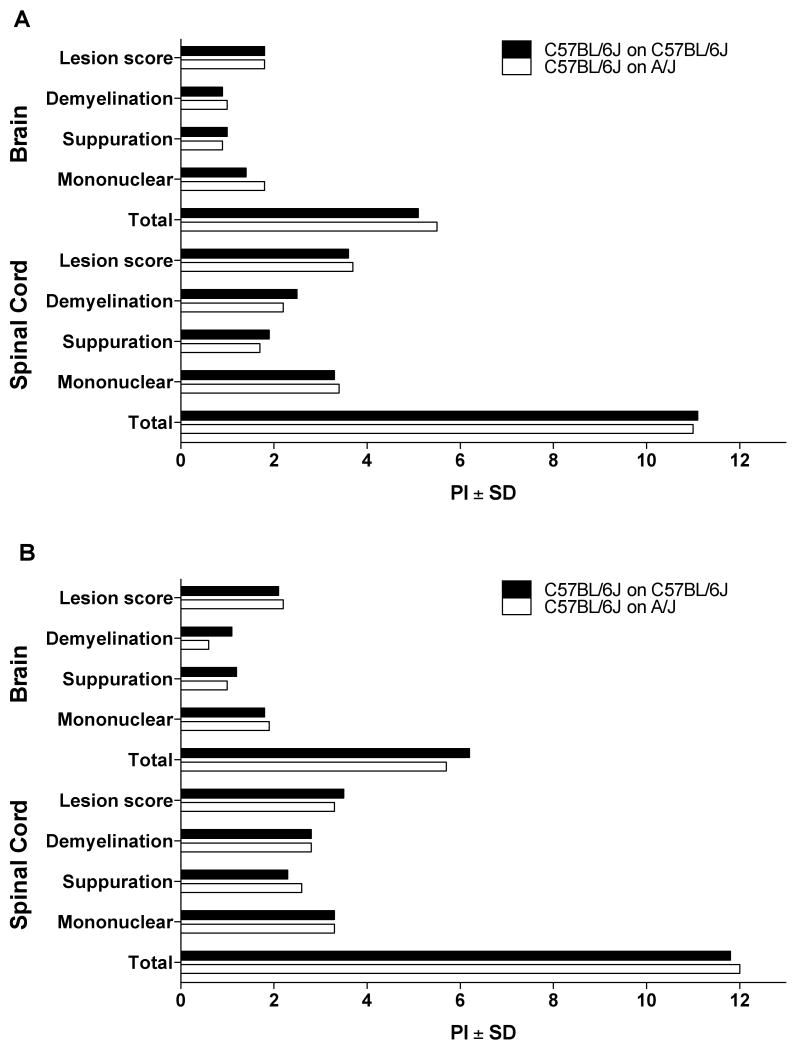
Figure 1.
Quantification of EAE pathology in natural and cross-fostered B6 female mice. EAE pathology in the brains and spinal cords of natural and cross-fostered mice elicited by immunization with (A) 1× MOG35–55+CFA+PTX and (B) 2× MOG35–55+CFA was evaluated in a semiquantitative manner. Lesions in the brains and spinal cords of natural and cross-fostered mice elicited with both protocols were not significantly different. The significance of differences was determined using the Mann Whitney test with a significance threshold of p = 0.05.

