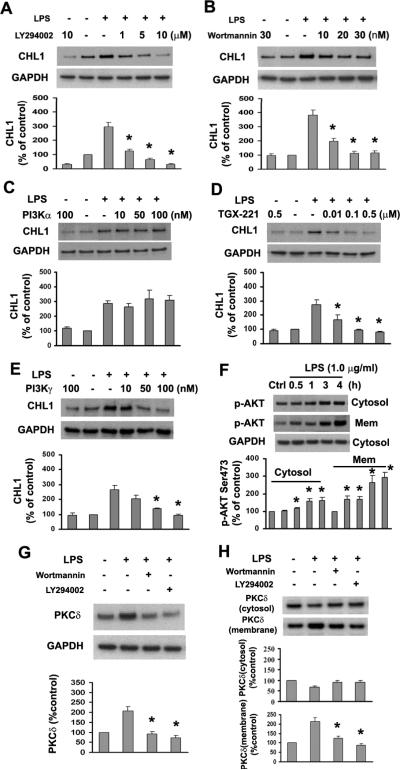
Fig. 5.
Inhibition of PI3K reduces LPS-induced CHL1 and PKCδ expression in cultured astrocytes. A–E. Expression of CHL1 protein was determined by immunoblot analysis in cells pretreated with LY294002, Wortmannin, or inhibitors for PI3Kα, β, γ for 1 h, followed by incubation with or without 1.0 μg/ml LPS for 24 h. *P<0.05 versus LPS alone group. F. Time-dependence of Akt phosphorylation (Ser473) in the subcellular fractions upon treatment with 1.0 μg/ml LPS. *P<0.05 versus control group without LPS treatment. G. Whole-cell lysates were subjected to immunoblot analysis for PKCδ. H. Cytosolic and membrane fractions were analyzed by immunoblot for PKCδ in astrocytes treated with LY294002 (10 μM) or Wortmannin (30 nM) for 1 h before incubation with 1.0 μg/ml LPS for 30 min.

