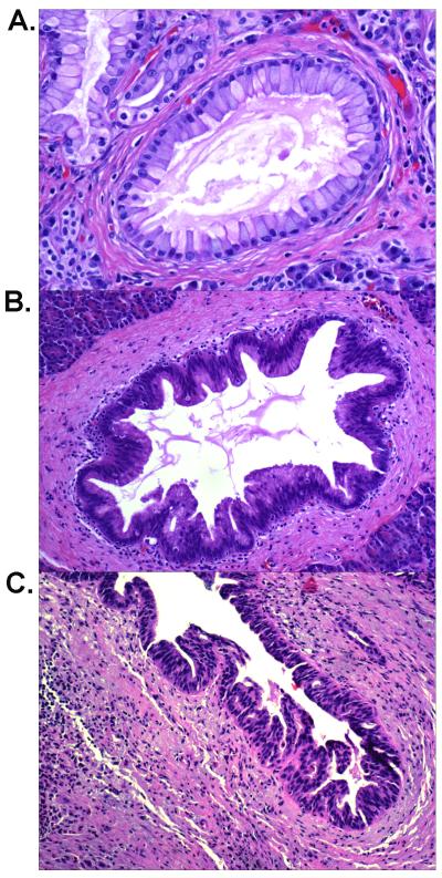
Figure 1.
Representative hematoxylin-eosin stained PanIN lesions from case 51 of the familial group. A: A PanIN-1 lesion showing mucinous columnar epithelial proliferation with little nuclear atypia (200X); B: A PanIN-2 lesion showing proliferated ductal epithelium with some nuclear atypia and pseudostratification (100X). C: A PanIN-3 lesion showing marked architectural and nuclear atypia (100X).

