
FIGURE 2.
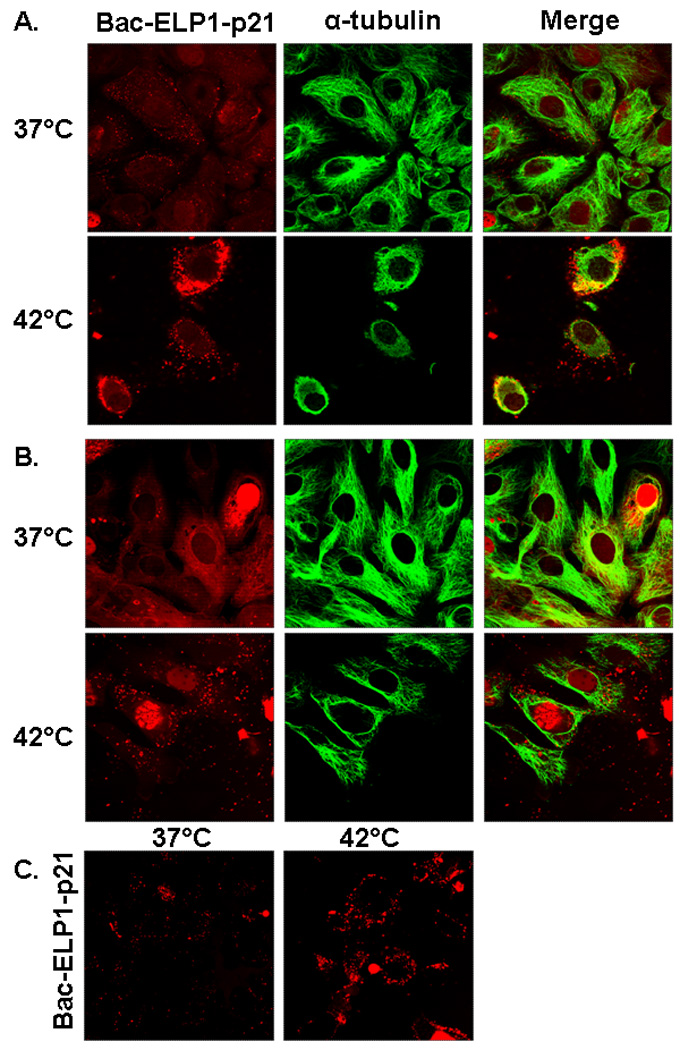
Subcellular localization of rhodamine labeled Bac-ELP1-p21 in SKOV-3 cells as visualized by confocal microscopy. Cells were treated with 20 µM of rhodamine-labeled Bac-ELP1-p21 at 37°C or 42°C for 1 h. Confocal images were taken (A) 1 h and (B) 24 h later. Tubulin was stained as a reference for cellular structure. The subcellular distribution of Bac-ELP1-p21 was also confirmed in live cells 24 h after a 1 h exposure at 37 or 42 °C (C). Fluorescence intensity in the heated samples was greater and the gain was adjusted for these samples before image acquisition. Therefore, these images are not quantitative.