
Fig.4. Slug is expressed in an AP1-dependent manner.
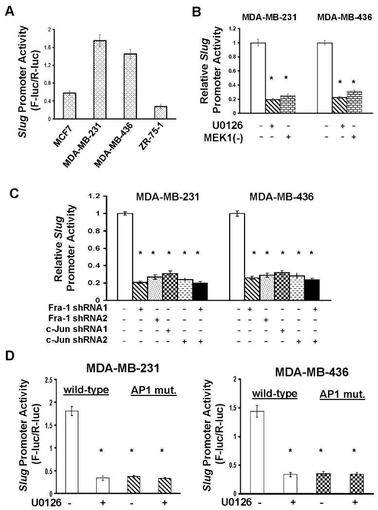
A. MCF7, MDA-MB-231, MDA-MB-436 and ZR-75-1 cells were transfected with the slug promoter luciferase reporter plasmid for 2 days, then lysed and cell lysates measured for luciferase activity as described in “Materials and Methods”. Data are means ± S.E. B. MDA-MB-231 and MDA-MB-436 cells were transfected with the slug promoter plasmid for 1 day and 5μM U0126 then added to cells for another 2 days. In a parallel expreriment, the slug promoter plasmid was co-transfected into cells with dominant negative MEK1 expression vector [MEK1(-)] for 3 days. Cells were lysed and cell lysates analyzed for luciferase activity. Data are means ± S.E. (*, p < 0.005 vs control). C. The slug promoter plasmid was transfected into MDA-MB-231 and MDA-MB-436 cells that expressed Fra-1 or c-Jun shRNA or both for 2 days. Cells were lysed and cell lysates analyzed for luciferase activity. Data are means ± S.E. (*, p < 0.005 vs control). D. MDA-MB-231 and MDA-MB-436 cells were transfected with the slug promoter plasmid or plasmid containing slug promoter with mutation in AP1 consensus site for 1 day and 5μM U0126 added to cells for another 2 days. Cells were lysed and cell lysates measured for luciferase activity. Data are means ± S.E. (*, p < 0.005 vs cells with wild-type promoter/no U0126).