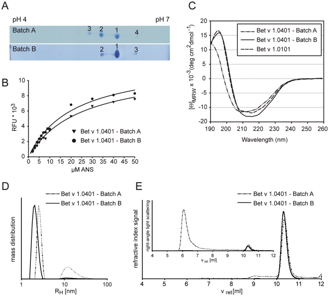
Figure 3. Physicochemical characterization of Bet v 1.0401.
Bet v 1.0401 batches were separated by 2D gel electophoresis. Gels were coomassie-stained, numbered spots were analyzed by MS and assigned to respective charge variants (A). ANS titration curves were recorded in 10 mM sodium phosphate (pH 7.4) using excitation and emission wavelengths of 370 and 450 nm, respectively. Solid lines represent nonlinear regressions of the experimental data (B). Circular dichroism spectra are presented as mean residue molar ellipticity [Θ]MRW at a given wavelength and baseline corrected. Bet v 1.0101 was used as reference for Bet v 1-like fold (C). Aggregation behaviour of Bet v 1.0401 batches was investigated by dynamic light scattering (DLS) in aqueous solution at a concentration of 1 mg/ml (D) and by online HPSEC-light scattering analysis operated at 0.5 ml/min in 0.1 M sodium phosphate pH 6.5, 150 mM sodium chloride (E). Analysis of Bet v 1.0401 batch A with DLS showed higher aggregation tendency (66% RH of 2.6±0.3 nm and 34% RH of 16±8 nm) compared to batch B (98% RH of 2.1±0.3 nm and 2% RH of 16±10 nm) (D). In online HPSEC-light scattering of batch A, molecular weight values of 70–80 kDa and 17 kDa were determined from refractive index and right-angle light scattering signals from peaks of oligomeric and monomeric Bet v 1.0401 eluting at retention times (vret) of 9.1 and 10.3 ml, respectively. For batch B one peak at vret of 10.3 ml with a molecular weight of 17 kDa was detected.

