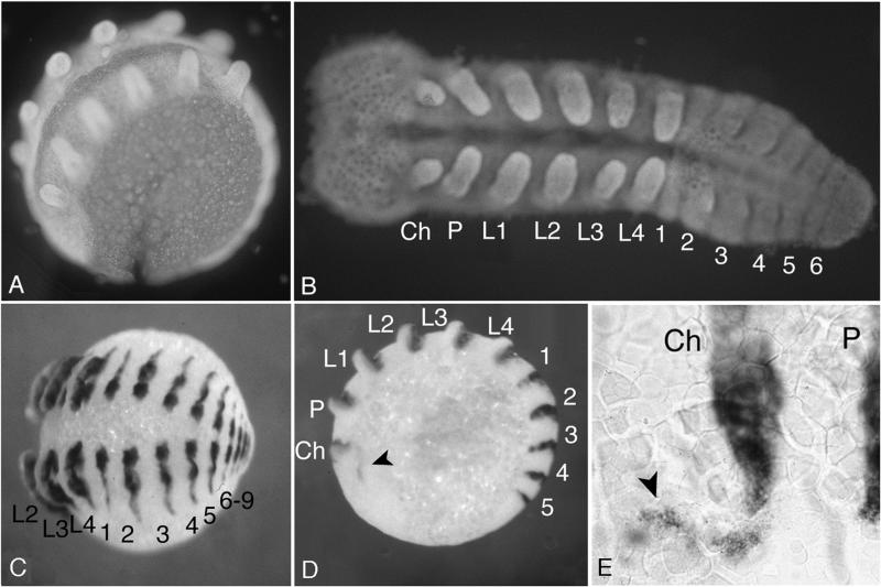
Figure 2.
Embryos of C. salei stained with 4′,6-diamidino-2-phenylindole (A and B) and with the EN probe by whole-mount in situ hybridization (C–E). Embryos were stained at midstage embryogenesis, when their general appearance is most reminiscent of the phylotypic stage of arthropods. (A) Embryo within the egg. (B) Embryo dissected free from yolk and flattened out. The anterior segments with their appendages already have formed fully while the abdominal segments are still incomplete. (C) Whole embryo stained with the EN probe. This embryo is at a later stage than the one in B and shows the final number of abdominal segments. (D) Embryo at an earlier stage showing an additional expression of EN in the head region (marked with an arrowhead), which might correspond to the preantennal segment. (E) Enlargement of the region showing the preantennal EN expression. Ch, cheliceres; P, pedipalps; L1–L4, leg 1–leg 4; 1–9, abdominal segments 1–9.

