Figure 1.

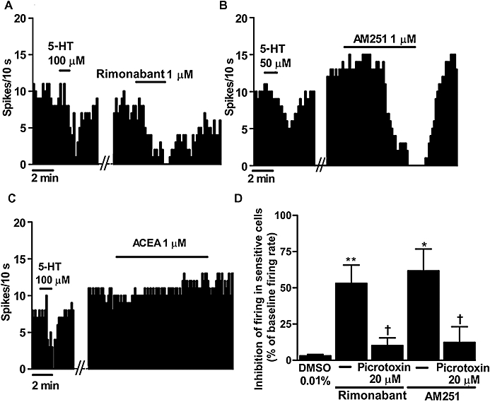
(A,B) Representative examples of firing rate recordings from three dorsal raphe nucleus (DRN) 5-hydroxytryptamine (5-HT) neurons, which show the inhibition of the firing activity of these neurons by the CB1 receptor antagonists SR141716A (rimonabant) (1 µM) (A) or N-(piperidin-1-yl)-5-(4-iodophenyl)-1-(2,4-dichlorophenyl)-4-methyl-1H-pyrazole-3-carboxamide (AM251) (1 µM) (B) and the slight increase of the 5-HT cell firing activity by the selective CB1 receptor agonist arachidonoyl-2-chloroethylamide (ACEA) (1 µM) (C). The vertical lines refer to the integrated firing rate values (spikes per 10 s), and the horizontal lines represent the time scale. Drugs were superfused at the concentration and for the time indicated by the horizontal bars; ACEA was applied in the continuous presence of the vehicle. (D) Bar histograms showing the inhibition of the firing activity of sensitive cells (mean ± SEM) from slices superfused with the vehicle (n= 5), rimonabant (1 µM) (n= 9), rimonabant (1 µM) + picrotoxin (20 µM) (n= 5), AM251 (1 µM) (n= 6) and AM251 (1 µM) + picrotoxin (20 µM) (n= 3). *P < 0.05, **P < 0.005 compared with the vehicle [dimethyl sulphoxide (DMSO)] group, and †P < 0.05 compared with the corresponding value in the absence of picrotoxin by Student's t-test. Note that both CB1 receptor antagonists decrease the firing activity of DRN 5-HT cells, which is blocked by picrotoxin.
