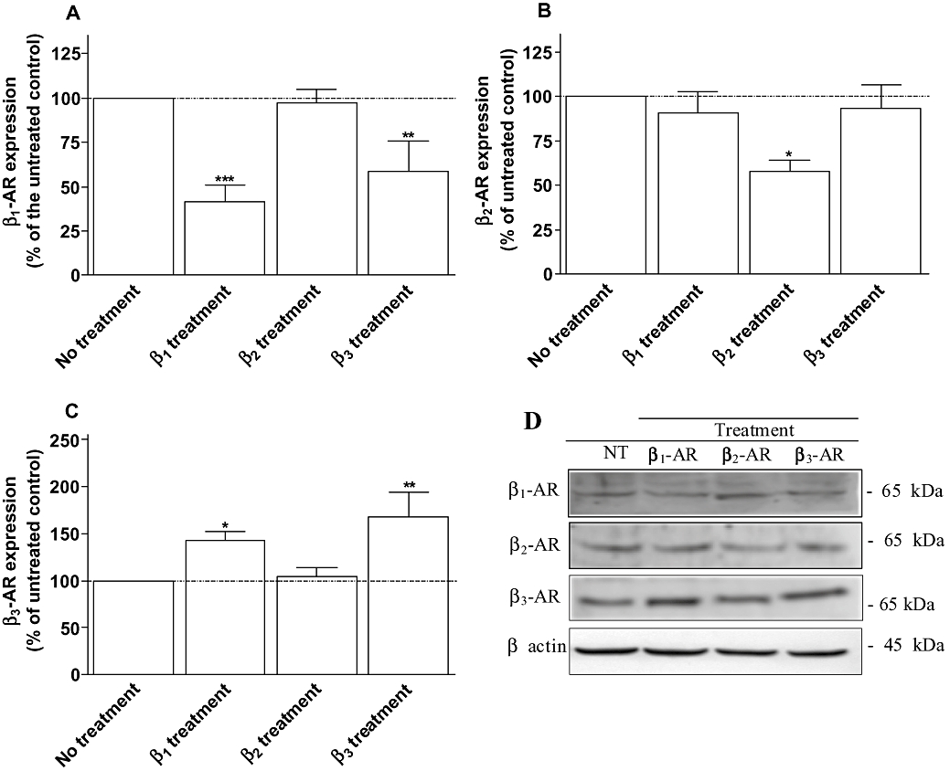
Figure 3.

Expression of β1- (A), β2- (B) and β3-adrenoceptors (C) obtained from neonatal rat cardiomyocytes untreated (control), treated with 10 µM dobutamine in the presence of 1 µM ICI 118551 (β1 treatment), 10 µM procaterol in the presence of 1 µM CGP 20712A (β2 treatment) or 2 µM CL-316243 (β3 treatment) for 24 h. Cell membrane homogenates were monitored by Western blotting for β1-, β2- and β3-adrenoceptors as described under Methods. The same samples were also analysed on separate blots using an antibody that recognizes β actin to confirm equal loading on each lane. Representative immunoblots for each proteins are shown in panel C. Data are expressed as the percentage of untreated cardiomyocytes (100%) following the calculation of β-adrenoceptor/β-actin ratio for each lane. The combined results (panel A, B and C) obtained from densitometric analysis of blots represent the mean ± SEM of five to seven independent experiments. *P < 0.05 and **P < 0.01 versus untreated control.
