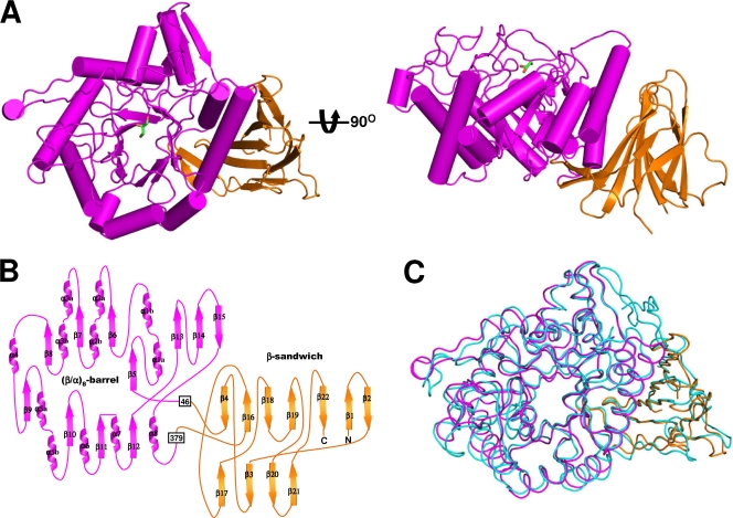
FIG. 2.
Overall shape of SrfJ. (A) A cartoon of the overall fold of SrfJ. The figure at right is rotated 90° in a vertical direction relative to the left-hand figure. The (β/α)8-barrel and the β-sandwich domains are shown in magenta and orange, respectively. A glycerol molecule bound in SrfJ is shown in a stick model (green). (B) Two-dimensional topology of SrfJ. Secondary structure elements of SrfJ are labeled appropriately and shown with the same color scheme as in panel A. (C) Structural superposition of SrfJ with human GlcCerase (PDB code 2v3e). The (β/α)8-barrel and the β-sandwich domains of SrfJ are shown in the same color scheme as in panel A, and human GlcCerase is depicted in light cyan.

