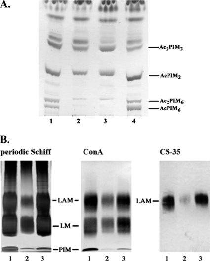
FIG. 4.
Analysis of polar PIMs LM and LAM from WT M. tuberculosis H37Rv, the H37RvΔRv3779 mutant, and H37RvΔRv3779/pVV16-Rv3779. (A) Equal amounts of total cellular lipids from WT H37Rv (lane 1), the Rv3779 mutant H37RvΔRv3779 (lane 2), the mutant carrying an empty plasmid, H37RvΔRv3779/pVV16 (lane 3), and the complemented mutant H37RvΔRv3779/pVV16-Rv3779 (lane 4) were analyzed by TLC developed in CHCl3-CH3OH-H2O-NH4OH (65:25:4:0.5). (B) LM and LAM extracted from equal-weight cells of WT H37Rv (lane 1), the Rv3779 mutant H37RvΔRv3779 (lane 2), and the complemented mutant H37RvΔRv3779/pVV16-Rv3779 (lane 3) were separated on a 10 to 20% Tricine gel and revealed by periodic acid-Schiff staining. The Western blot analyses were performed on the same samples using concanavalin A (ConA) reacting with the t-Manp residues of LM/LAM, as well as the CS-35 monoclonal antibody known to react with the arabinan segment of LAM.

