Figure 1.
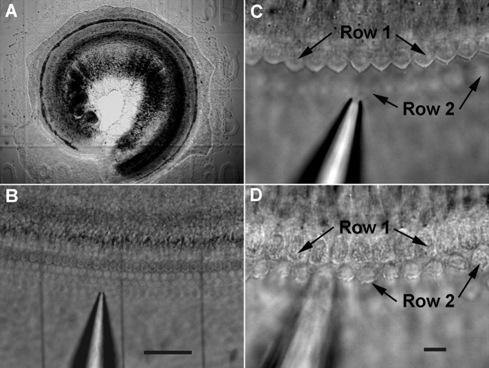
Images of the tissue culture of neonatal organ of Corti and procedures to remove the hair bundles. A, Survey microphotograph of a 1-d-old culture of basilar membrane—organ of Corti prepared from the basal turn of a neonatal gerbil cochlea. The culture shown in this picture was explanted on the gridded coverslip. B, Three glass fibers with diameter of ∼5–6 μm were inserted underneath the culture to mark the area where the hair bundles were removed and the area used for control. C, The hair bundles observed under high magnification before the bundles were ablated. The bundles in the first row of OHCs were in focus. A suction pipette was positioned nearby. D, The hair bundles in first row OHCs were removed by the suction pipette. The bundles of the second row of OHCs were damaged. The focus in D was slightly readjusted to show the bundleless reticular lamina in the first row of OHCs and the damaged hair bundles in the second row of OHCs. Scale bars: B, 50 μm; C, D, 10 μm.

