
Figure 2.
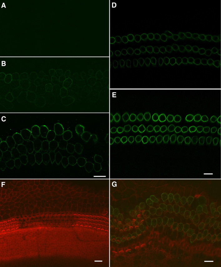
Confocal images of prestin expression in the OHCs. A, One day after the hair bundles were removed. No prestin immunoactivity was seen at this age. B, C, Six and eleven days after the bundles were removed. Prestin was expressed at 6 PID and strongly expressed at 11 PID. Scale bar: 10 μm for A–C. D, E, Prestin expression in the preparations isolated from the developing gerbils at 7 (D) and 12 (E) d after birth. Both were from middle turns. F, G, Hair bundles and prestin expression at 1 PID. The hair bundle was labeled with rhodamine-phalloidin. Three rows of “V”-shaped OHC bundles and one row of IHC bundles could clearly be seen in the control areas. The hair bundles were no longer present in the area (middle of the image) where the hair bundles were removed. No prestin immunoactivity was seen at this age. G, Hair bundles and prestin expression at 11 PID. The hair cell region expanded significantly at this age. Prestin immunoactivity could be seen in the OHCs with and without the hair bundles.