
Figure 4.
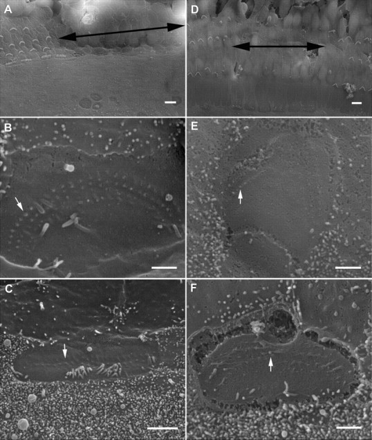
SEM pictures of cultured organ of Corti and hair cell stereocilia. A, Surface view of the reticular lamina at 7 PID. The area where the bundle was removed is marked by double-head arrows. The tissue was from the middle turn. No regrowth or elongation of the truncated stereocilia was seen. B, Apical surface of a bundleless OHC under high magnification. The rootlets of three rows of truncated stereocilia were clearly visible. A white arrow indicates one of the rootlets. A kinocilium is still present in the apical surface. C, A bundleless IHC. Some microvilli together with a kinocilium are also seen. B and C are both from the damaged area from A. D, Surface view of the reticular lamina at 13 PID. No bundle repair was observed. The tissue was from basal turn. E, F, High-magnification images of the apical surface of an OHC (E) and IHC (F). Arrows mark the rootlets of stereocilia. E and F are from the damaged area shown in D. Scale bars in A and D represent 10 μm, while the bars in the rest of the images represent 1 μm.