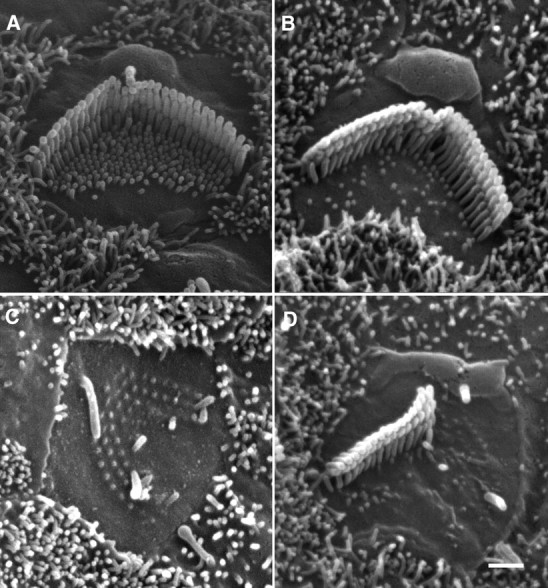
Figure 5.

SEM picture of hair cell stereocilia. A, Stereocilia of a 2-d-old OHC in culture. Kinocilium and abundant microvilli are clearly visible at this age. B, Stereocilia of a 14-d-old OHC in culture. The kinocilium disappears and the number of microvilli is significantly reduced at this age. C, Apical surface of an OHC at 7 PID. The kinocilium remains while no elongation of the stereocilia is seen. D, Apical surface of an OHC at 7 PID. Half of the stereocilia bundle is undamaged and the kinocilium is also present. No repair is seen on the truncated stereocilia.
