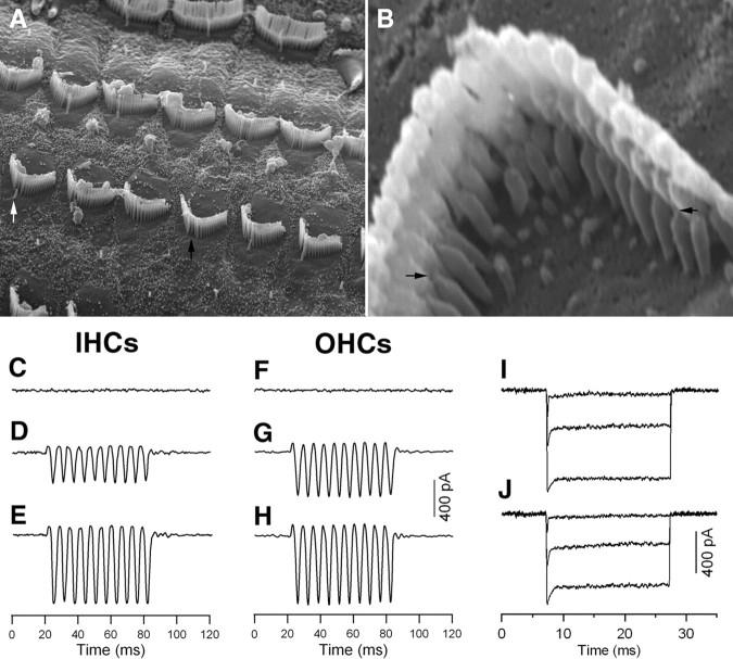
Figure 6.
SEM picture of hair cell stereocilia and the MET currents measured from cultured hair cells. All cultures were prepared from the cochlear apical turn from 8-d-old gerbils for all experiments. A, SEM of stereociliary bundles after cultured for 24 h. At this age, the kinocilium in some hair cells already disappeared (indicated by a black arrow) and in other hair cells, the kinocilium was still present (marked by a white arrow). B, Newly regenerated tip links 24 h after being cut. Tip links could clearly be seen in the apex of the innermost (shortest) stereocilia to the side of their taller neighbor. Two black arrows mark the two examples of the newly regenerated tip link. C, F, MET current recorded 10 min after BAPTA treatment. E, F, MET current recorded from control IHC and OHC after 24 h culture. D, G, MET current returned in the BAPTA-treated preparations after 24 h culture. The cells were held at −70 mV. The bundle was vibrated in response to fluid jet stimulation at 100 Hz. Three presentations were averaged for each trial. E, H, MET currents measured from cultured hair cells whose tip links were not cut (control group). I, J, Adaptation of MET currents measured from control (I) and BAPTA-treated (J) OHCs after 24 h culturing. The hair bundle was deflected by a rigid fiber which was driven by piezoelectric actuator. Three presentations were averaged for each trial.

