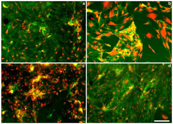
Fig. 2.
Fluorescence microscopy of lectin- and anti-CD31 antibody stained human (HUVEC, a, c) and mouse (lung b, d) endothelial cells. a, b cells were stained with Cy5.5-labeled tomato (L. esculentum), lectin (at 5 μg/ml, shown in red) and Alexa Fluor 488-labeled CD31 monoclonal antibody (1 μg/ml, shown in green); c, d cells were stained with Cy5.5-labeled U. europaeus lectin (L. esculentum), lectin (5 μg/ml, shown in red) and Alexa Fluor 488-labeled CD31 monoclonal antibody (1 μg/ml, shown in green). Anti-CD31 antibodies were species-specific. Bar=50 μm.

