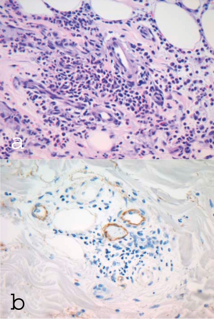
Figure 2.
Dermal histology and immunohistology in hypocomplementemic urticarial vasculitis syndrome (HUVS)
a) Dermal perivascular mixed-cell inflammatory cell infiltrate, predominantly granulocytes, with destruction of small vessels and nuclear dust; original magnification ×400
b) Immunohistochemical staining for C1q: demonstration of C1q deposits on the vascular endothelium

