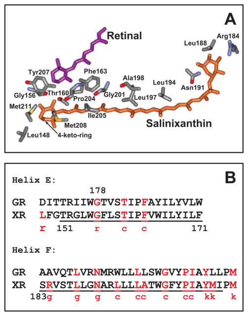
Figure 1.
A. Conformation of salinixanthin and residues within 4 Å of salinixanthin in xanthorhodopsin, based on the 3DLL coordinate set (20). The acyl tail of the carotenoid is in contact with lipids, and is not shown. The distances between the side chains and carotenoid are presented in Supporting Information, section I. B. Conservation of residues that interact with the carotenoid in xanthorhodopsin in gloeobacter rhodopsin. Letters c, g, k, and r stand for the chain, glucoside, keto-group, and ring, respectively, and indicate part of the carotenoid in the vicinity of a residue.

