Figure 3.
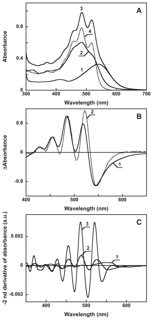
A. Absorption spectra of: 1, gloeobacter rhodopsin, 16 μ in 0.02% DDM, pH 7.2, 100 mM NaCl, 4 mm pathlength; 2, 8 μ salinixanthin in the same buffer; 3, mixture after adding 8 μ of salinixanthin to 16 μ to the gloeobacter rhodopsin; 4, spectrum 3 minus spectrum 1. B. Absorption changes of salinixanthin upon binding to: 1, gloeobacter rhodopsin (difference between spectrum 4 and 2 in panel A); 2, xanthorhodopsin (taken from (23), Figure 6A, curve 4). C. Second derivatives of absorption spectra 1 through 3 in Figure 3A (multiplied by minus 1).

