Figure 1.
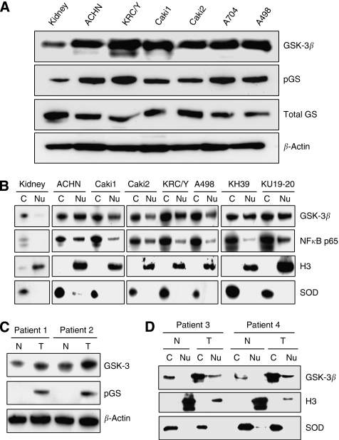
GSK-3β is overexpressed in nuclei of renal cancer cells. (A) Protein lysates from the indicated RCC cell lines and normal kidney as a control were separated by SDS–PAGE (50 μg per well), transferred to PVDF membrane and probed with antibodies against GSK-3β, phospho-glycogen synthase (pGS) and total glycogen synthase (GS). (B) Cytosolic (C) and nuclear (Nu) fractions were prepared from RCC cell lines and normal kidney, separated by SDS–PAGE (50 μg per well), transferred to PVDF membrane and probed with indicated antibodies. Cu/Zn supeoxide dismutase (SOD) and histone H3 (H3) were used as cytosolic and nuclear markers, respectively. (C) Expression of GSK-3β and pGS was detected in protein extracts from primary tumour (T) and corresponding normal kidney tissue (N) obtained from kidney cancer patients. (D) Nuclear (Nu) and cytosolic (C) fractions were prepared from fresh tumour (T) and corresponding normal kidney tissue (N) sampled from kidney cancer patients, and analysed as described in (B).

