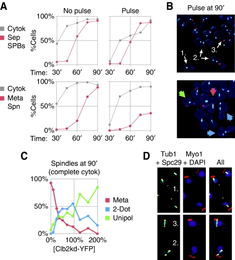
Figure 3.
Persistent Clb2 after mitotic exit causes aberrant spindle assembly in the next cell cycle. (A) SPB duplication and separation (red, top, BD144c-8B) or metaphase spindle formation (red, bottom, BD127-11B) are plotted with completion of cytokinesis (gray) versus time post-release from cdc20 arrest for an unpulsed control and a low Clb2kd YFP pulse (70% peak at the beginning of the experiment, diluting to 40% peak by the end). (B) To the right of each graph are fluorescence microscopy images of cells 90 min post-release: Myo1–mCherry merged with Spc29–CFP (top) or CFP–Tub1 (bottom). For Spc29–CFP, cells with one, two, or three SPBs are indicated (arrows). For CFP–Tub1, arrows label cells with bar-shaped metaphase spindles (red), unipolar spindles (green) or short discontinuous ‘2-dot' spindles (blue). (C) Spindle morphology versus Clb2kd–YFP concentration among cells that have completed cytokinesis at 90 min post-release. Phenotypes charted are described in (B, bottom). (D) Cells bearing CFP–Tub1 and Spc29–YFP (YL178-1-12-2) were pulsed with untagged Clb2kd and released for 90 min from cdc20 arrest. Three spindle types are shown: (1) One dim SPB distant from DAPI staining that does not colocalize with tubulin (top), (2) two SPBs with discontinuous tubulin arrays (bottom), or (3) a normal bipolar spindle (bottom).

