Fig. 5.
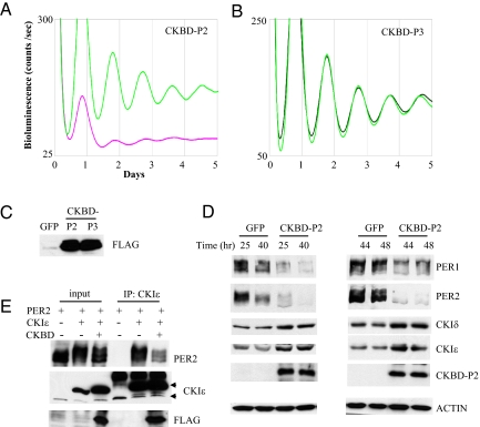
Disruption of PER interaction with CKIδ/ε by CKBD-P2 abolishes circadian rhythms and destabilizes PER in MEFs. (A and B) Casein kinase binding domain from PER2 (CKBD-P2) (A) and the corresponding domain from PER3 (CKBD-P3) (B) were overexpressed in wt MEFs and bioluminescence rhythms were measured as above. The FLAG tag added to the N-termini of both CKBDs was used to ensure similar expression levels. Note that the basal line for CKBD-P2 is extremely low. This was consistently observed in several experiments. (C) MEFs from (A) and (B) were immunoblotted for FLAG. (D) GFP and CKBD-P2 MEFs at indicated times were harvested and the extracts were immunoblotted. Note that endogenous CKIδ/ε levels were increased in CKBD-P2 MEFs. A dark exposure for PER2 blot in the left panels is shown in Fig. S7A to demonstrate that low levels in CKBD-P2 MEFs are not due to smearing of PER2 band. Blots are representative of three experiments. (E) PER2, CKIε and/or CKBD-P2 were overexpressed in wt MEFs, the cells were harvested 24 h after the infection and the extracts were subjected to IP for CKIε. The resulting immunocomplexes were immunoblotted for PER2, CKIε, and FLAG. Top and bottom arrow indicate exogenous and endogenous CKIε, respectively. Blots are representative of four experiments.

