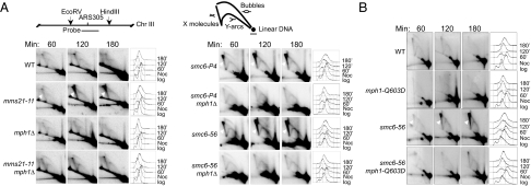
Fig. 4.
mph1Δ and mph1-Q603D decrease the levels of recombination intermediates in smc6 and mms21 mutants. (A and B) Cells were arrested at G2/M phase with nocodazole at 25 °C and then were released into YPD medium with 0.033% MMS at 30 °C. The replication and recombination intermediates at the ARS305 region 60, 120, and 180 min after release were analyzed by 2D gel electrophoresis followed by Southern blotting. Diagrams indicating the position of the probe and the replication structures are shown above the 2D gel images. The X-shaped DNA structures are indicated by arrowheads in smc6 and mms21 mutants. FACS analyses are presented to the right of each gel image.

