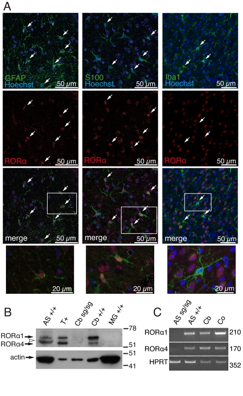
Fig. 1.
Expression of RORα in astrocytes. (A) Localization of RORα in brain. Sagittal sections of cerebellum (Left) and cortex (Middle and Right) from 21-d-old Rora+/+ mice. Astrocytes were labeled using anti-GFAP (green) or anti-S100 (green) antibodies, microglia using anti-Iba-1 (green), and nuclei using Hoechst 33258 (blue). Expression of RORα was revealed with anti-RORα (red). RORα and nucleus co-localize in astrocytes but not in microglia (arrows). Higher-magnification images of framed areas are in the merged images (Bottom). (B) Western blot analysis of RORα in enriched nuclear extracts from WT (+/+) and staggerer (sg/sg) cerebellum (Cb) and from astrocyte (AS) and microglia (MG) cultures. Twelve micrograms of tissue and 25 μg of cell extracts were electrophoresed. T+ are in vitro-synthesized RORα1 and RORα4 isoforms. The arrowhead indicates protein cross-reacting with anti-RORα antibody. (C) Amplification of RORα1 and RORα4 transcripts in cultivated astrocytes. Total RNA from highly purified staggerer (sg/sg) and WT (+/+) astrocyte cultures (AS) and from WT cortex (Co) and WT cerebellum (Cb) as positive controls were used. Predicted sizes for the amplified fragment are indicated (Right).

