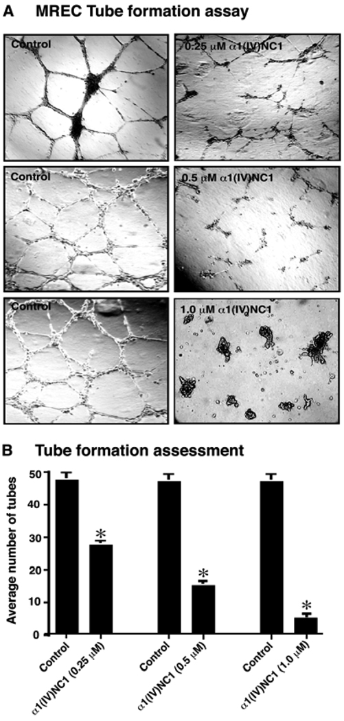
Figure 3.
(A) Tube formation assay. MREC were plated on Matrigel coated plates in retinal endothelial cell medium as control or with 0.25–1.0µM α1(IV)NC1 protein. Tube formation was evaluated using a light microscope, and representative fields at 100x magnification are shown. (B) Tube formation assessments. Graph displays the average number of tubes in 2 different wells in each condition and mean±standard error of the mean [SEM] of three independent experiments (n=6). *P<0.001 compared to control.

