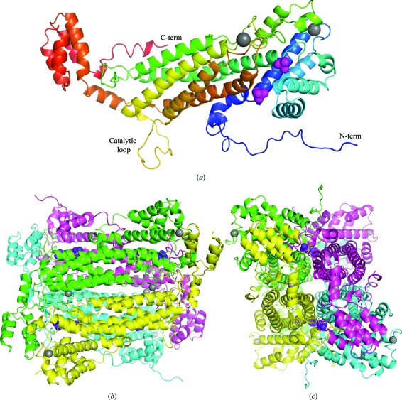
Figure 1.
(a) Crystal structure of a single subunit of adenylosuccinate lyase (ASL) from E. coli. The protein is coloured from the N-terminus (blue) to the C-terminus (red). The N- and C-termini and the catalytic loop are labelled. The phosphate (magenta) and sodium (grey) ions are shown as space-filling representations. (b) Structure of the tetramer of E. coli ASL. Two orthogonal views are shown. Figures were produced with PyMOL (http://pymol.sourceforge.net/).

