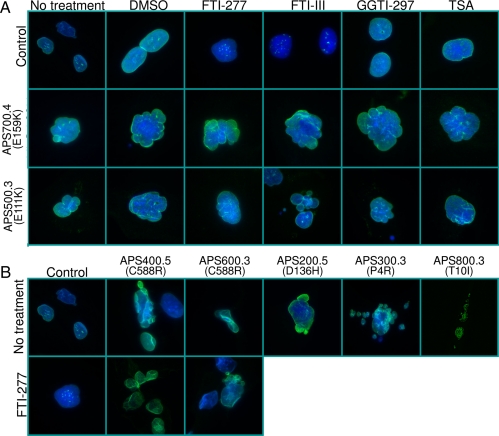
Figure 7.
Nuclear morphology of skin fibroblasts from APS patients and controls and response to various pharmacological agents. A, Indirect immunofluorescence microscopy was conducted using lamin A/C antibody (H-110). The top row shows control cells, the middle row shows cells from patient APS 700.4 (E159K), and the bottom row shows cells from patient 500.3 (E111K). The first column shows cell morphology without any treatment, the second column with dimethylsulfoxide, and the next columns after incubation of the cells for 48 h with 10 μm farnesyl transferase inhibitors, FTI-277 or FTI-III, geranylgeranyl transferase inhibitor, GGTI-297, or a histone deacetylase inhibitor, trichostatin A (TSA), respectively. Fibroblasts from both patients showed misshapen, bilobed, or multilobed nuclei or nuclear blebs. The localization of lamin A to the periphery of the nucleus was not affected. Incubation of the cells with various pharmacological agents failed to correct the nuclear abnormalities. B, Indirect immunofluorescence microscopy lamin A/C antibody (H-110) in control fibroblast cells and those from patients APS 400.5 (C588R), 600.3 (C588R), 200.5 (D136H), 300.3 (P4R), and 800.3 (T10I). The bottom row shows control cells and those from patients APS 400.5 (C588R) and 600.3 (C588R) after incubation for 48 h with 10 μm FTI-277.

