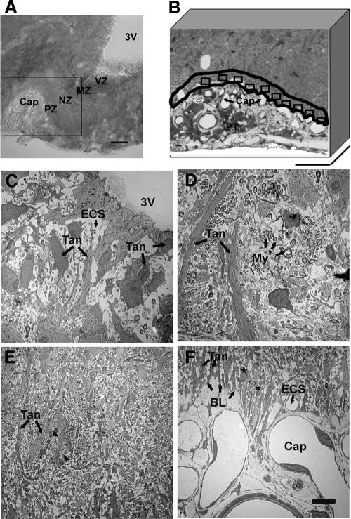
Figure 2.
Light and electron microscopic images of the lateral median eminence. A, Transmitted light differential interference contrast image shows the lateral median eminence from a representative young vehicle-treated rat. The lateral median eminence can be visually delineated into zones beginning dorsomedially with the third ventricle (3V), followed by the myelinated axon zone (MZ), the neuronal profile zone (NZ), the pericapillary zone (PZ), and finally the portal capillary vasculature (Cap). The area outlined in the box was chosen for subsequent electron microscopy analyses. Scale bar, 100 μm. B, The pericapillary zone in this semithin section (500 μm in thickness) was traced along the portal capillary basal lamina, and a 10-μm boundary was drawn (outlined in black). Ten subregions (rectangles) along the basal lamina were chosen for further electron microscopic analyses. The pars tuberalis (PT) can also be seen. Scale bar, 100 μm. C–F, Electron microscopic images show the characteristic features of the four zones of the lateral median eminence of a representative vehicle-treated rat. C, In the ventricular zone (VZ; A), tanycyte cell bodies (Tan; arrow) are seen, some of which loosely line the walls of the third ventricle. D, In the myelinated axon zone (MZ; A), groups of axons covered with myelin sheaths (My; arrows) are perpendicular to tanycyte processes (Tan; arrow). E, In the neural profile zone (NZ; A), axon swellings (arrowheads; note that these are more apparent at higher magnification) are in close proximity to the tanycyte processes (Tan; arrows). F, In the pericapillary zone (PZ; A), neuroterminals (asterisks) are separated by tanycytic endfeet (Tan; arrows) from the basal lamina (BL; arrows) of the portal capillaries (Cap). The extracellular space (ECS; arrow) surrounding neuronal and glial elements is clearly visible in the ventricular and pericapillary zone. Scale bar (C–F), 5 μm.

